
Varicose Veins
Varicose veins are one of the most common manifestations of chronic venous disease. They are most often caused by venous incompetence, which is a failure of the one-way valves inside the leg veins that normally keep blood flowing back up to the heart. When those valves don’t close properly, blood falls back down the leg with gravity, pressure inside the vein rises, and the vein becomes engorged, dilated and visible under the skin.
The experience of varicose veins is genuinely a spectrum. For some people, varicose veins are a cosmetic concern, taking the form of small, fine blue “spider veins” or larger, twisted, ropey veins sitting just beneath the surface. For others, they cause aching, heaviness and swelling through the day. At the more serious end, prolonged venous hypertension can lead to skin staining, lipodermatosclerosis, and venous leg ulcers that are slow to heal and frequently recur. Australia has one of the higher rates in the world: varicose veins affect roughly 10–20 per 1,000 people over a lifetime, and up to 25% in high-risk groups. If you’re troubled by them, you are far from alone.
Dr Irina Baimatova is a Perth-based vascular surgeon who manages the full spectrum of chronic venous disease. Her approach combines a comprehensive duplex ultrasound assessment at the time of your first appointment (so you leave with a clear diagnosis and a tailored plan), conservative management where appropriate, and a complete range of office-based and surgical treatments. Those treatments include endovenous laser ablation (EVLA), the VenaSeal non-thermal closure system, ultrasound-guided sclerotherapy, micro-sclerotherapy for spider veins, and open surgical stripping and avulsions when the surgical option is the right one for you.
What happens with varicose veins over time?
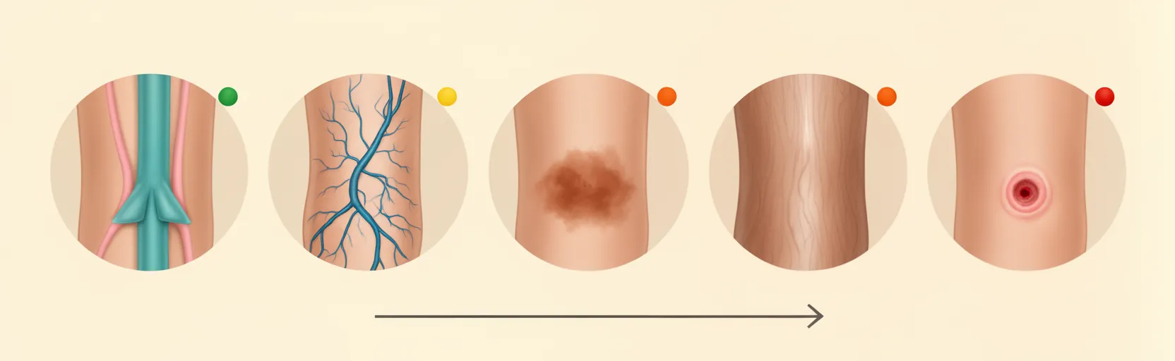
Chronic venous disease is progressive. The early signs are usually easy to live with; the late signs can be disabling. Knowing where you are on the spectrum is what makes the difference.
Cosmesis
Spider veins and visible varicose veins
Most patients first notice varicose veins as a visual change. They may appear as small, fine blue lines (often called spider veins or telangiectasias) or as larger, twisted, ropey veins sitting just beneath the surface of the skin. They are most commonly seen on the calves and the inside of the thighs, and they often become more prominent with prolonged standing, in warm weather, or as the day goes on.
For some patients, the cosmetic change is the main concern. For others, it is a marker that something is starting to go wrong underneath.
Skin Changes
Haemosiderin Staining and Lipodermatosclerosis
When blood pools in the veins, the pressure inside them rises. Red blood cells can then leak out into the surrounding tissue, and the iron they contain (haemosiderin) stains the skin. This is most often seen around the ankles, where the skin becomes a darker brown or deep red. The affected skin looks shiny, feels firmer than normal, and is increasingly prone to injury and ulceration.
Lipodermatosclerosis is a more advanced stage of skin change. It results from chronic inflammation of the skin and underlying fat, leading to thickening, hardening, and a woody texture. Once this is established, the skin is fragile, and even small cuts and scratches can become problematic.
Swelling and Oedema
As venous pressure rises, fluid leaks out of the veins into the surrounding tissue. The result is ankle and lower-leg swelling that worsens through the day and is often worst by evening. Many patients find the swelling is worse on days spent standing or sitting, and improves overnight when the legs are elevated. Compression stockings typically help significantly in the early stages.
Ulceration
Once skin changes begin, the skin becomes vulnerable. Small cuts or scratches can fail to heal, become infected, and develop into venous leg ulcers. They usually appear on the medial (inner) side of the lower leg (the so-called gaiter area). Although they often start small, they can enlarge rapidly. Venous ulcers are typically shallow, may ooze, and are often painful. Even after they heal, the recurrence rate is high unless the underlying venous reflux is treated.
This is the stage of the disease at which treatment is most urgent, and where modern treatment has the biggest impact. The EVRA trial showed that early venous reflux ablation, in addition to compression bandaging, significantly speeds ulcer healing and reduces recurrence.
Superficial Venous Thrombosis (SVT)
A superficial venous thrombosis is a clot in a varicose or superficial vein. It most often presents as a painful, red, tender lump or cord running along the line of a varicose vein, classically along the great saphenous vein (GSV) on the inner thigh or calf. The redness is inflammation, not infection, so antibiotics are not required.
A duplex ultrasound is used to confirm the diagnosis, exclude a concurrent deep vein thrombosis (DVT), and tell us how close the clot sits to the deep venous system. An SVT within 3 cm of the junction with a deep vein, or longer than 5 cm, is treated with anticoagulation for at least 45 days (3 months for higher-risk patients). An SVT within 3 cm of the deep system is treated as a DVT, with full therapeutic anticoagulation.
What to be Aware of with Varicose Veins
The underlying mechanism: valvular incompetence and venous reflux
Healthy leg veins contain one-way valves that open as blood is pushed upwards by the calf muscle pump, then close to stop gravity pulling it back down. When those valves fail (become incompetent), blood falls back down the leg with each pump cycle. This is called venous reflux.
Over time, the reflux causes the affected vein to dilate and become tortuous, and the pressure inside it rises. That pressure is called venous hypertension. The downstream effect is the spectrum of symptoms described above: swelling, skin staining, lipodermatosclerosis, and eventually ulceration.
The calf muscles are the engine that returns blood to the heart, so anything that weakens them (including inactivity and obesity) makes the situation worse.
Where Reflux Occurs
Venous reflux can occur in any of the veins of the lower limb:
- Great saphenous vein (GSV): the most common site, running along the inside of the thigh and calf.
- Small saphenous vein (SSV): running up the back of the calf.
- Accessory saphenous veins.
- Perforator veins: normally carry blood from superficial to deep veins (the “re-entry” flow). When they become incompetent they can cause local varicose veins and contribute to disease progression.
- Pelvic veins: pelvic vein incompetence is increasingly recognised as a contributor, particularly in women with recurrent or atypical varicose vein patterns.
A complete duplex ultrasound is needed to map all of these and to plan treatment.
Risk factors
Some risk factors you can modify, others you cannot. The most important are:
- Obesity, which increases venous pressure and reduces calf muscle pump efficiency
- Age, as valve function declines with age
- Previous DVT, which can damage valves directly
- Family history of varicose veins, a strong heritable component
- Previous leg trauma
- Prolonged standing or sitting: occupations like nursing, teaching, hairdressing, retail and driving are over-represented
- Physical inactivity, which weakens the calf muscle pump
- Pregnancy and oral contraceptive use, as varicose veins frequently appear or worsen during pregnancy, and may persist afterwards
- High blood pressure
- Smoking
Effective treatments for varicose veins (Pros and Cons)
Treatment is individualised. The right choice depends on the anatomy of your veins (mapped on ultrasound), the severity of your symptoms, your general health, your preferences, and what is covered by your health fund.

Conservative Management
Conservative measures are the foundation of treatment, and they continue alongside any procedural option.
Elevation can significantly reduce swelling in the short term. Exercise and weight loss help by improving the calf muscle pump and reducing the pressure on the veins. Compression therapy, usually class II compression stockings at 20–30 mmHg, fitted with adequate arterial supply, prevents blood from pooling, reduces the backward flow of blood, and has been shown to slow the long-term progression of skin changes. Compression is also the cornerstone of venous ulcer care, in conjunction with the treatments below.
Endovenous laser ablation (EVLA)
Thermal
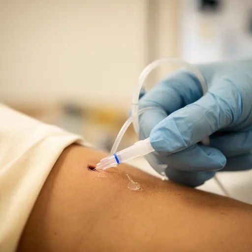
A laser fibre is placed into the incompetent vein under ultrasound guidance. The laser energy heats the vein from the inside, causing it to close down on itself. Blood is then redirected into the surrounding competent deep veins, relieving the pressure and the symptoms it caused.
EVLA is usually performed as a day procedure in the rooms under local anaesthetic (tumescence) and is often not fully covered by private health funds. A partial Medicare rebate is usually available. The tumescent anaesthetic acts as both a pain block and a heat sink to protect the surrounding tissues, and ultrasound guidance allows us to close as much of the vein as safely as possible while protecting the deep system. The smaller, more tortuous tributaries are typically treated with ultrasound-guided sclerotherapy at the same time, which gives the best cosmetic result. Spider veins are treated with micro-injection sclerotherapy.
VenaSeal
non-thermal
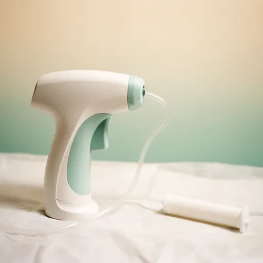
VenaSeal is a medical-grade adhesive (glue) that closes the incompetent vein mechanically rather than with heat. Because it is non-thermal, it does not require tumescent anaesthetic, which means significantly fewer needle injections (a real benefit for needle-phobic patients), and no risk of heat-related nerve injury. Outcomes are comparable to thermal ablation.
Small tributary veins are still treated with ultrasound-guided sclerotherapy, and spider veins with micro-sclerotherapy.
Open surgical stripping and avulsions
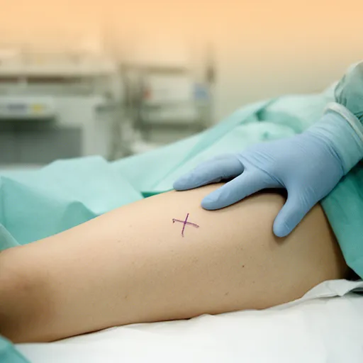
Open surgery is covered by most private health funds with vascular cover. It involves a small incision in the groin to disconnect the great saphenous vein from the deep femoral vein, passing a thin plastic “stripper” down the vein, and removing it through a small incision near the knee. The smaller varicose veins are removed via tiny 2–3 mm incisions (avulsions) and closed with steri-strips or glue.
Open surgery is performed in hospital under general anaesthetic. The groin incision takes about a week to heal. Recovery is typically 1–2 weeks off work and a little more discomfort than the percutaneous (in-rooms) options, but it remains a reliable, well-established option for patients whose anatomy is not suited to the catheter-based techniques, or who prefer the surgical route.

Recurrence
Recurrence after any varicose vein treatment is roughly 10% at 10 years. With thermal ablation and surgical stripping, true re-canalisation of the treated vein is rare. What is more common is neovascularisation, the formation of new veins in the same area, and the progression of disease in veins that were previously normal. Both are manageable, often with a straightforward secondary procedure.
Information for GPs
What to do before referring
A trial of conservative management is appropriate in the first instance for most patients. This includes leg elevation, regular exercise, weight optimisation, and class II compression stockings (20–30 mmHg), provided arterial supply is adequate (check ABPI in older patients, diabetics, or anyone with PAD risk factors). Most patients do not need pre-referral imaging, as the duplex ultrasound is performed in the rooms at the first specialist appointment. A more complete picture can be taken in a single visit.
Compression stocking prescribing
Class II compression (20–30 mmHg) is the standard for chronic venous disease. Always check arterial supply (ABPI) before prescribing compression in older patients, in diabetics, and in anyone with PAD risk factors. Compression is contraindicated in critical limb ischaemia, decompensated heart failure, and acute cellulitis. For patients who struggle with knee-high stockings, thigh-high or panty-style compression is usually available.
How to refer
- Phone: (08) 6333 2866
- Fax: (08) 9467 6227
- Email: admin@drbaimatova.com.au
- Healthlink EDI: ibaimato
- Post: PO Box 124, West Perth WA 6872
* For complex cases or to discuss a patient before referral, the rooms can be contacted on the phone number above.
Recognising red flags
- Venous ulceration: almost always in the medial gaiter area. Refer early, particularly in conjunction with active compression bandaging.
- Haemosiderin staining and lipodermatosclerosis: these are pre-ulcer states. Treatment of the underlying reflux can prevent progression.
- SVT close to the deep system junction: within 3 cm of the saphenofemoral or saphenopopliteal junction, treat as a DVT with full therapeutic anticoagulation.
- Long-segment SVT (>5 cm in length): treat with 45 days of anticoagulation (3 months for higher-risk patients).
- Sudden painful cord-like lump along a varicose vein: assume SVT until proven otherwise. Confirm with duplex and exclude concurrent DVT.
SVT management summary
Confirm with duplex ultrasound and exclude concurrent DVT. Treat with anticoagulation for 45 days (low-risk short distal SVT) up to 3 months (higher-risk, including SVT within 3 cm of the deep system, which should be treated as a DVT with full therapeutic anticoagulation). Antibiotics are NOT indicated, as the erythema is inflammatory, not infectious.
Where care is delivered
- Consulting rooms: Hollywood Medical Centre (85 Monash Avenue, Nedlands WA 6009); St John of God Midland.
- Operating: Hollywood Private Hospital, St John of God Subiaco, St John of God Midland, Royal Perth Hospital, St John of God Midland Public.
- Telehealth: available on request.
When to refer
Symptomatic varicose veins
Aaching, heaviness, throbbing, night cramps, or persistent swelling that has not responded to conservative measures in primary care
Skin changes
Haemosiderin staining, lipodermatosclerosis, atrophie blanche, or venous eczema indicating chronic venous insufficiency that warrants specialist review.
venous ulceration
Active or recurrent venous ulcers; early referral for endovenous ablation alongside compression bandaging is supported by the EVRA trial.
Superficial venous thrombosis
SVT located within 5 cm of the saphenofemoral or saphenopopliteal junction, or any thrombosed superficial segment measuring more than 5 cm.
Varicose veins in pregnancy
Symptomatic veins persisting or worsening 3–6 months postpartum, where conservative measures have failed and treatment is being considered.
Cosmetic concerns
Symptomatic veins persisting or worsening 3–6 months postpartum, where conservative measures have failed and treatment is being considered.
Failed conservative management
Ongoing symptoms or progressive venous disease despite an adequate trial of lifestyle advice and compression therapy.
Frequently Asked Questions
Q. What are varicose veins, and how are they different from spider veins?
Varicose veins are dilated, tortuous, ropey veins that sit just under the surface of the skin, usually on the legs. They are caused by incompetent valves in the underlying veins, leading to venous reflux and high pressure inside the vein. Spider veins (also called telangiectasias or thread veins) are the much finer, web-like blue or red lines that sit within the skin itself. They are usually cosmetic, but they can sometimes indicate underlying venous reflux that should be assessed with ultrasound.
Q. Are varicose veins dangerous, or are they just cosmetic?
For some people they are purely cosmetic, and that is a perfectly valid reason to seek treatment. For others they are the visible sign of chronic venous disease, which can progress to swelling, skin staining, lipodermatosclerosis, painful superficial venous thrombosis, and ultimately venous leg ulcers. Treating the underlying reflux early can prevent the later stages of the disease.
Q. What causes varicose veins, and am I at risk?
The main cause is incompetent venous valves (venous reflux), usually in the great or small saphenous vein. Risk factors include a family history of varicose veins, age, obesity, pregnancy, occupations that involve prolonged standing or sitting, previous DVT, smoking, and physical inactivity. A duplex ultrasound is the standard test to confirm the cause and map the anatomy.
Q. Do I need a referral to see a vascular surgeon for varicose veins?
A GP referral is required for Medicare to rebate part of the specialist consultation. Your GP can also help coordinate pre-referral conservative care (compression, weight, exercise) and provide the clinical history that helps us plan.
Q. What is a venous duplex ultrasound, and do I need one?
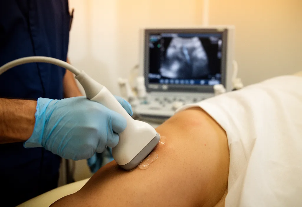
A duplex ultrasound is a non-invasive scan that uses sound waves to visualise the veins of your legs in real time, including the direction of blood flow. It is the gold standard for diagnosing varicose veins and planning treatment. It is performed in the rooms at your first appointment, so you leave with a complete picture in a single visit.
Q. Which varicose vein treatment is best: laser, VenaSeal, sclerotherapy, or surgery?
There is no single “best” treatment. The right option depends on the anatomy of your veins (which the duplex ultrasound maps), the severity of your disease, your general health, and your preferences. In broad terms, endovenous laser and VenaSeal are the modern first-line options for axial vein reflux; ultrasound-guided sclerotherapy treats the tortuous tributary veins; micro-sclerotherapy treats spider veins; and open surgery remains a reliable option when catheter-based techniques are not suitable or when the surgical route is preferred. Dr. Baimatova will discuss all of these with you after the ultrasound.
Q. Is varicose vein treatment covered by Medicare or private health insurance?
Medicare provides a partial rebate for the consultation and for the ultrasound. For the procedural options, endovenous laser and VenaSeal performed in the rooms are often not fully covered by private health funds, although a Medicare rebate usually applies. Open surgical stripping and avulsions performed in hospital are generally covered by private health funds with vascular cover, subject to your policy’s excess and waiting periods. Our team can help you clarify your cover before booking.
Q. How long is recovery after endovenous laser ablation or VenaSeal?
Most patients return to normal light activity within 1–2 days and to full activity, including exercise, within 1–2 weeks. Compression stockings are usually worn for 1–2 weeks after the procedure. Some bruising and tenderness along the treated vein is normal and resolves over a few weeks.
Q. Will my varicose veins come back after treatment?
Recurrence after any varicose vein treatment is approximately 10% at 10 years. The most common cause is neovascularisation, the formation of new veins in the treated area, or progression of disease in previously normal veins. If recurrence occurs, a straightforward secondary procedure is usually effective.
Q. Can varicose veins cause leg ulcers, and can the ulcers be treated?
Yes. Venous leg ulcers are the most serious complication of chronic venous disease, and they are most often caused by the same underlying reflux that causes varicose veins. Modern care combines compression bandaging with early venous reflux ablation. The EVRA trial showed that early ablation significantly speeds ulcer healing and reduces recurrence. Dr. Baimatova manages venous ulcers in conjunction with your GP and wound-care nurse.
Q. What is a superficial venous thrombosis (SVT), and when does it need anticoagulation?
A superficial venous thrombosis is a clot in a varicose or superficial vein, presenting as a painful, red, tender cord along the vein. The redness is inflammation, not infection, so antibiotics are not needed. A duplex ultrasound confirms the diagnosis and tells us how close the clot sits to the deep system. An SVT longer than 5 cm, or within 3 cm of the junction with a deep vein, is treated with anticoagulation for at least 45 days (3 months in higher-risk patients). An SVT within 3 cm of the deep system junction is treated as a DVT.
Q. I have varicose veins during pregnancy. What can I safely do?
Varicose veins frequently appear or worsen during pregnancy and may improve in the months after delivery. Conservative measures such as leg elevation, exercise, and class I or II compression stockings (20–30 mmHg) are the mainstay during pregnancy and breastfeeding. Most procedural treatments are deferred until at least 3–6 months postpartum so the veins have a chance to settle. If the veins remain symptomatic after that, a full assessment and treatment plan can be made.
Q. Do you offer consultations in the rooms, and is telehealth available?
Yes to both. Initial consultations are generally in the rooms at Hollywood Medical Centre (Nedlands) or St John of God Midland, where the duplex ultrasound can be performed on the day. Telehealth consultations are available on request for review appointments, country patients, and patients who find it difficult to attend in person.
1. Geroulakos, G., & Paraskevas, K. I. (2024). The 2024 ESVS Guidelines on Lower Limb Peripheral Arterial Disease: A Step Forward. European Journal of Vascular and Endovascular Surgery, 67(1), 3-5
2. Lampridou, S., Rawasdheh, M., Saghdaoui, L. B., Wells, M., & Davies, A. H. (2024). Factors affecting adherence to medication, smoking cessation and exercise in patients with peripheral artery disease. JVS-Vascular Insights, 100074.
3. Nordanstig, J., Behrendt, C. A., Baumgartner, I., Belch, J., Bäck, M., Fitridge, R., … & ESVS Guidelines Committee. (2024). Editor’s Choice–European Society for Vascular Surgery (ESVS) 2024 Clinical Practice Guidelines on the Management of Asymptomatic Lower Limb Peripheral Arterial Disease and Intermittent Claudication. European Journal of Vascular and Endovascular Surgery, 67(1), 9-96.
4. Ten Berg, J., Sibbing, D., Rocca, B., Van Belle, E., Chevalier, B., Collet, J. P., … & Mehilli, J. (2021). Management of antithrombotic therapy in patients undergoing transcatheter aortic valve implantation: a consensus document of the ESC Working Group on Thrombosis and the European Association of Percutaneous Cardiovascular Interventions (EAPCI), in collaboration with the ESC Council on Valvular Heart Disease. European heart journal, 42(23), 2265-2269.
5. Leng, G. C., Fowler, B., & Ernst, E. (2000). Exercise for intermittent claudication. The Cochrane database of systematic reviews, (2), CD000990. https://doi.org/10.1002/14651858.CD000990
6. Harwood, A. E., Pymer, S., Ingle, L., Doherty, P., Chetter, I. C., Parmenter, B., Askew, C. D., & Tew, G. A. (2020). Exercise training for intermittent claudication: a narrative review and summary of guidelines for practitioners. BMJ open sport & exercise medicine, 6(1), e000897. https://doi.org/10.1136/bmjsem-2020-000897
7. Guidon, M. (2011). An exercise programme for patients with intermittent claudication: randomised trial of health outcomes and cost analysis (Doctoral dissertation, Royal College of Surgeons in Ireland).